 Faster, More Comfortable Mammograms
Now available at Three Rivers Hospital — Waverly, TN — (931) 296-0223
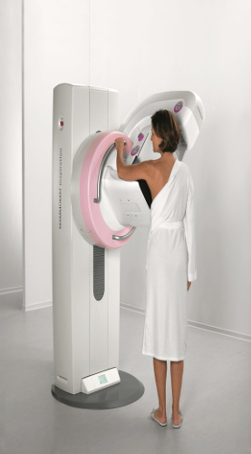
Siemens Digital Mammography System Provides High Quality Breast Imaging in a Patient-Friendly Environment
Three Rivers Hospital has a new digital mammography system that can provide a more pleasant, soothing exam environment for patients while helping to minimize radiation dose and provide physicians with vital information. The MAMMOMAT® Inspiration from Siemens Healthcare captures breast images with an X-ray detector that converts images into a digital picture that can be displayed immediately on the system's computer monitor.
Unlike film, digital mammograms allow physicians to manipulate image characteristics such as magnification, orientation, brightness and contrast, which improves the ability to view specific areas of the breast. With the MAMMOMAT Inspiration system, results are produced more quickly, helping to decrease anxiety for patients.
To help minimize the patients’s radiation dose and exposure time during a mammogram, the Inspiration automatically selects the appropriate dose for individual patients’ breast characteristics. To help further ensure patient comfort, the Inspiration applies compression only as long as the patient’s breast is soft and pliable.
Early detection through regular mammograms is the key to diagnosing and beating breast cancer. The MAMMOMAT Inspiration digital mammography system offers a faster exam that minimizes compression time on the breast, resulting in a less painful experience for you. And the outstanding image quality can help your doctor identify breast cancer at the earliest possible stage, using the lowest possible radiation dose.
* One in eight women is diagnosed with breast cancer each year in America, which is the most common form of cancer in women. Mammography can help detect breast tumors at their earliest, most treatable stages.
What is a Mammogram
A mammogram is an X-ray image of your breast used to screen for breast cancer. Mammograms play a key role in early breast cancer detection and helps decrease breast cancer deaths.
During a mammogram, your breast are compressed between two firm surfaces in order to spread out the breast tissue. Then an X-ray captures black-and-white images of your breast that a doctor uses to detect changes and cancer.
A mammogram can be used either for screening or for diagnostic purposes. How often you should have a mammogram depends on your age and your risk of breast cancer.
Why its done
Mammography is an X-ray imaging of your breast designed to detect tumors and other abnormalities. Mammography can be used either for screening or for diagnostic purposes in evaluating a breast lump.
Screening mammography
Screening mammography is used to detect breast changes in women who have no signs or symptoms or observable breast abnormalities. The goal is to detect cancer before clinical signs are noticeable. A screening mammogram consist of two mammogram image of each breast taken from different angles.
Diagnostic mammogram
Diagnostic mammography is used to investigate suspicious breast changes, such as a breast lump, breast pain, an unusual skin appearance, nipple thickening, or nipple discharge. It is also used to to evaluate abnormal findings on a screening mammogram A diagnostic mammogram includes additional mammogram images, such as magnification and compression views.
When to begin screening mammography
Experts and medical organizations do not agree on when women should begin regular mammograms or how often the test should be performed. Talk to your doctor about the risk factors, your preferences, and the benefits and risks of screening. Together, you can decide what screening mammography schedule is best for you.
Some general guidelines for when to begin screening mammography include:
Women with a normal risk of breast cancer
Many women begin mammograms at age 40 and have them every one to two years.
Women with a high risk of breast cancer
Women with a high risk of breast cancer may benefit by beginning screening mammograms before the age of 40. Talk to your doctor for an individualized program.
Risks
Mammograms expose you to low dose radiation. The dose is very low, and fore most women the benefits of regular mammograms outweigh the risk posed by this amount of radiation.
How you prepare
Bring your prior mammogram images if they were done at a different facility so that the radiologist can compare with your new images.
Avoid using deodorants, antiperspirants, powders, lotions, creams, or perfumes under your arms or on your breast. Metallic particles in powders and deodorants could be visible on your mammogram and cause confusion.
What you can expect
You will be given a gown and asked to remove neck jewelry and clothing from the waist up. To make it easier wear a two piece outfit that day.
For the procedure itself, you will stand in front of an X-ray machine specially designed for mammography. The technician places one of your breast on a platform and raises or lowers the platform to match your height, The technician will help you position your head, arms, and torso to allow an unobstructed view of your breast.
Your breast is gradually pressed against the platform by a clear plastic plate. Pressure is applied for a few seconds to spread out the breast tissue. The procedure is not harmful, but you may find it uncomfortable.
Your breast must be compressed to even out its thickness and permit the X-ray to penetrate the breast tissue. The pressure also holds your breast still to decrease blurring from movement and minimizes the dose of radiation needed. During the brief X-ray exposure, you will be asked to stand still and hold your breath.
After images are made of both of your breast, you may be asked to wait while the technician checks the quality of the images. If the views are inadequate for technical reasons, you may have to repeat part of the test. The entire procedure usually takes less than 30 minutes. Afterward you may get dressed and continue normal activities.
Results
The Radiologist views and interprets the results and will send a final report to your doctor, who will then explain the results to you.

